Assignment 1:
The Sea Star Tube Feet!
 |
| Figure 1: Sea Star tube feet retrieved from http://www.gettyimages.ca/photos/tube-feet?sort=mostpopular&excludenudity=true&mediatype=photography&phrase=tube%20feet Sea Stars are echinoderms that are related to sand dollars, sea urchins, and sea cucumbers. They live among a variety of ocean floors, including tropical and cold habitats, as well as in brackish waters. Sea stars live do not live in freshwater environments as they require salt water for survival. The five-arm sea stars are the most common but some species can have up to 10, 20, and even 40 arms! Sea stars have bony, calcified skin that protects them from most predators, and come in a variety of wear striking colors that camouflage them or scare off potential attackers. |
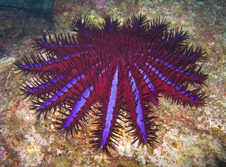 |
| Figure 2: Sea Star with many legs retrieved from https://en.wikipedia.org/wiki/Starfish |
Surprisingly, the sea star is a carnivore, as they normally feed on clams or oysters. The way sea stars feed is remarkable. They are able to consume prey outside their bodies by expelling their sac-like cardiac stomach from their mouth onto their prey. The stomach will then envelop the prey to digest it and finally withdraws back into the body. The sea stars open shells of the oysters and clams through the use of their tube feet on the bottom of their arms.
Fun Facts:
Sea stars have no brain and no blood. Their nervous system is spread through their arms and their “blood” is actually filtered sea water called hemolymph.
Also, sea stars have eye spots at the end of each arm. It is a very simple eye that does not see much detail but can sense light and dark.
 |
| Figure 3: Retrieved from https://s-media-cache-ak0.pinimg.com /736x/06/75/e1/0675e138049703e4a2353fd18968db21.jpg |
Starfish can change their gender depending on the temperature of the ocean or the availability of food. Usually, sea stars are born male and produce sperm, then part way through their life they transform into females and produce eggs. In fact, one female can have up to 1.3 million eggs.
Structure
 |
| Figure 4: Sea Star arms anatomy retrieved from http://tolweb.org/Asteroidea
Sea star tube feet are essential for locomotion, feeding, and respiration. They have hundreds of these tube feet and they are located on their underside. Tube feet are short, hollow, elastic, thin-walled, closed tubes that extend through a gap, called ambulacral pore, which lies between two ambulacral ossicles.The radial canal runs down the entire length of the sea star arm and receives water from the annular, which is then passed into the tube feet.
|
 |
| Figure 5:Structure of the Sea Star tube foot retrieved from https://www.britannica.com/science/tube-foot |
Tube feet consist of three parts, the ampulla, the podia, and the sucker. Ampulla, are rounded sac-like structures situated above the ambulacral ossicle that projects into the coelom and containing both circular muscles and longitudinal muscles. When these muscles contract it allows water to enter into the tube foot, allowing it to extend, and when it dilates the foot retracts. The podia are the middle tubular portion extending through the ambulacral groove and are covered externally by ciliated epithelium and internally with peritoneum. Between these two layers lie connective tissue in a crossed-fiber helical array and longitudinal muscle fibers. The fiber angle of the connective tissue allows protraction and prevents dilation of the tube feet, it also limits elongation of the ampullae. Lastly, the sucker is at the lower end of the podium and is flatted forming a cup-like structure.
The tube feet epithelium is covered by a thin cuticle that is continuous with the covering of adjacent areas of the ambulacrum. When the tube feet are retracted, the epithelium is thick and the epithelial surface is highly folded into annular rings. When tube feet are protracted, the epithelium will appear thinner and the folding will be reduced.
 |
| Figure 6: Starfish tube feet sec, 7um H&E Microscope Slide Retrieved from http://www.carolina.com/animal-microscope-slides/starfish-tube-feet-sec-7-um-h-e-microscope-slide/308218.pr |
The distal conical end of the tube feet have an epithelium made up of tall columnar cells with intensely staining cell inclusions. Underneath the epithelium layer lies nervous tissue containing many nerves and neurons. Below the nervous tissue, there is a dense layer of fibrous connective tissue with the fibers arranged in a cross-helical array. The connective tissue is of great importance for the mechanism of the tube feet as it reinforces the wall so that an increase in pressure causes an increase in length, rather than diameter preventing large swelling or stretching. Below the dense connective tissue sheet, there is a robust layer of muscle fibers which are arranged in circumferential bands around the lumen if the ampullae. The internal lumen of the tube feet is also lined with an epithelial layer.
The radial canal as previously mentioned runs down the entire length of the sea star arm and receives water from the annular, which is then passed into the tube feet. The connective tissue of the ambulacrum will surround the radial canal preventing it from expanding. There are no muscle fibers encircling the radial canal.
 |
| Figure 7: Reproduced from McCurley et, al. 1995. Biology Bulletin |
Mechanism of Locomotion
 |
| Figure 8: Seastar using its tube feet to maneuver across the sand retrieved from http://0.tqn.com/d/marinelife/1/0/2/5/-/-/seastar-tubefeet- wildcatdunny-flickr-500x375.jpg |
Here is a short video of the sea star's tube feet at work!
https://www.youtube.com/watch?v=T1pQe9dWXuQ
Function
Tube feet serve many functions to the sea stars survival. They are extremely helpful in locomotion as previously mentioned. Without the use of the tube feet, sea stars would not be able to maneuver around the ocean floors in the hunt for prey. The tube feet also help the sea star to burrow within the mud and sand by bending away from the ambulacrum toward the sides of the arm. The tube foot bends laterally into the sediment and then retracts and bends back toward the ambulacrum to repeat the cycle until the sea star is burrowed beneath the sediment.
 |
| Figure 9:Retrieved from http://disney.wikia.com/wiki/Peach |
The sea star tube feet are also helpful in feeding. They help the starfish to hold onto food or pry clams open. They are also responsible for the direct uptake of organic nutrient material directly across the tube feet epithelium due to the secretion of mucus and reabsorption. Lastly, the tube feet are in involved in conveying food down the length of the ambulacral groove to the mouth. As a particular food approaches a given tube foot, it bends toward the particle and protracts until the tip adheres to the food. Once attached, the tube foot retracts, pulling the food toward the mouth.
 |
| Figure 10: Seastar eating an anchovy using its tube feet. Retrieved from https://www.reddit.com/r/pics/comments/1xiigs/starfish_eating_an_anchovy/ |
References
Kennedy, J. (2016, April 25). 10 Facts About Starfish (Sea Stars). Retrieved from http://marinelife.about.com/od/invertebrates/tp/seastarfacts.htm
McCurley, R.S., Kier, M.W. (1995). The function morphology of starfish tube feet: The role of a crossed-fiber helical array in movement. Biology Bulletin, 188(2). 197-209. http://www.jstor.org.qe2a-proxy.mun.ca/stable/pdf/1542085.pdf
National Geographic. (n.d.). Starfish (Sea Star). Retrieved
from http://animals.nationalgeographic.com/animals/invertebrates/starfish/
Smith, J. E. (2009). The structure and function of the tube feet in certain echinoderms. Journal of the Marine Biological Association of the United Kingdom, 22(1), 345-357. Retrieved from https://www.cambridge.org/core/journals/journal-of-the-marine-biological-association-of-the-united-kingdom/article/the-structure-and-function-of-the-tube-feet-in-certain-echinoderms/4051CAB92EEDFCEC8CBF2F9A05E57BAA
Ursadhip. (2011, September 3). Tube Feet of Echinoderms. Make Easy Zoology. Retrieved from http://ursadhip.blogspot.ca/2011/09/tube-feet-of-echinoderms.html
Wikipedia The Free Encyclopedia. (2016, October 18). Tube Feet. Retrieved from https://en.wikipedia.org/wiki/Tube_feet